


 312 338

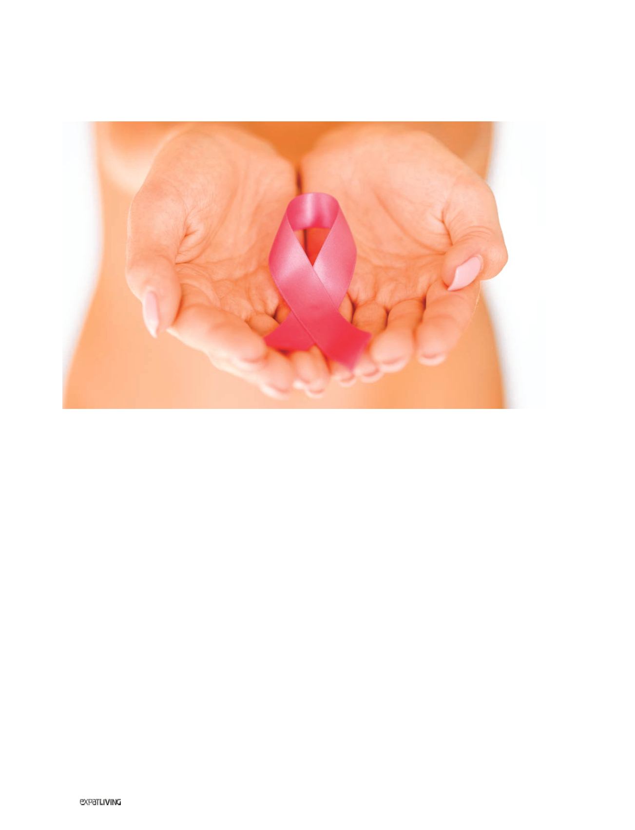
HEALTH&FITNESS
312
June14
need to examine the axillary (armpit)
lymph nodes – each of the 20 or more
nodes is about the size of a mung
bean – because that’s the first place the
breast cancer cells migrate to.”
HOW IT’S DONE:
“After injecting a blue
dye into the breast and massaging it, we
can then make just a small incision at
the armpit area in order to identify the
sentinel lymph node (SLN), the first one
that receives the lymphatic drainage. If
testing shows the SLN to be cancer-free,
we know that the cancer is in its early
stage, so there’s no need to remove any
other lymph nodes.
COMMENT:
“This advance makes a
big difference to the patient’s long-term
comfort. That’s because our lymphatic
system regulates fluid drainage from
the arms, and disrupting it through the
wholesale removal of lymph nodes can
leave you with a swollen arm or stiff
shoulder for life.”
#3 Radiation during surgery,
rather than at a later stage
“Radiation during surgery is a pet topic
of mine,” says Georgette, who has had
experience of performing this technique
at the European Institute of Oncology in
Milan, where it was first developed.
“The usual procedure,” she explains,
“is to apply radiation to the whole chest
wall, in repeated small doses over the
course of a month – after their breast-
conserving surgery, after the wound has
healed and after chemotherapy. So, for
three to four weeks, the patient has to
visit the radiation centre every day.
“But now, for selected cases – often
older patients with less aggressive
tumours – the radiation can be done
during the surgery itself, and that will
be the only radiation that’s required.”
HOW IT’S DONE:
“Within the operating
theatre, the surgeon uses a light-weight,
mobile radiation machine that emits
a beam of electrons. Depending on
the machine used, it can take from 10
minutes to 45 minutes to deliver the
required dose.
“The beauty of this is that one can
focus exactly on the affected location,
thus sparing other areas from the
effects of radiation. And it can be done
while the surgical team is waiting for the
pathology report. To calculate the dose
of radiation, a radiation physicist plus a
radiation oncologist are on hand, too,
so it can get pretty crowded in there!”
COMMENT:
“So far, there’s only one
such unit in Singapore so far, and it’s at
the National Cancer Centre.”
#4 Shrinking tumours
before surgery
Usually done by the medical oncologist,
this is also a very exciting development,
says Georgette. “It’s especially useful
for big tumours, where we don’t have
enough skin to close the mastectomy
wound, or if the patient is very keen to
have breast-conserving surgery.”
HOW IT’S DONE:
“After doing a biopsy,
we work with the medical oncologist to
tailor a chemotherapy that will shrink the
tumour to a more manageable size. In
the space of three to four months, after
four rounds of chemo, some tumours will
shrink to half their size; others shrink so
effectively that you can hardly feel them
through the skin. Occasionally, we have
to put a clip into the middle of the tumour
before starting chemotherapy, so as to
be able to find the tumour at the time
of surgery.”
COMMENT:
“Being able to shrink the
tumour in size can mean converting
the treatment from a full mastectomy to
breast-conserving surgery.”
Syda Productions | Dreamstime.com